MRI is a medical imaging technique that examines tissues, organs and skeletal system. Magnetic resonance imaging scanners create highly detailed two- and three-dimensional images of human anatomy using a strong magnetic field and radio waves. MRI is very common in the diagnosis and treatment of diseases of the brain, spine, joints and abdomen. Its major advantage compared to other imaging techniques is that it does not need ionizing radiation. MRI is a revolutionary technology, particularly in neurology and brain research.
Table of Contents
What is MRI?
Magnetic resonance imaging (MRI) creates cross-sectional images of organs and tissues in the body using magnetic field and computer generated radio waves. It produces high resolution images of body parts that cannot be imaged by X-ray and computed tomography or ultrasound. A MRI device is a magnet with an open tube at both ends.
The signal that forms the magnetic resonance image comes mostly from the nuclei in the body’s fat and water molecules. Magnetic field or radio waves are not felt during the scan, the process itself is painless.
What is an MRI scanner?
A regular MR unit is in the form of a large cylinder surrounded by a circular magnet. It has a desk that slides towards the center of the magnet. There is a possibility to have an Magnetic resonance imaging device open on its sides. The magnetic field produced by the MRI scanner produces images by temporarily rearranging the water molecule nuclei (protons) in the body.
At the center of each hydrogen atom that forms water molecules, there is a smaller particle called a proton. Protons are like tiny magnets and are very sensitive to magnetic fields. The magnetic resonance scanner temporarily realigns protons by creating a very strong magnetic field. When the magnetic field is closed, the protons gradually return to their normal alignment.
During this return, the protons generate a radio signal that is measured by the receivers in the scanner and converted into an image. These signals carry information about the location of protons in the body. They also help to distinguish between various tissue types in the body, because protons of different tissue types line up at different speeds and produce different signals.
This process does not cause chemical changes in the tissues. Scanner settings can be adjusted to create a contrast between different body tissues. Additional magnetic fields can produce 3D images that can be viewed from different angles. (1)
What is magnetic resonance spectroscopy?
Magnetic Resonance Spectroscopy (MR Spectroscopy – MRS) is a special MRI technique. It is an advanced MRI application that measures the biochemical changes of lesions detected in the brain and gives definitive diagnostic value.
It shows if there is a tumor in the brain by comparing the normal brain with the abnormal brain tissue to see and produces a detailed map of the chemical structure of the tumor. (2)
What does MRI do and why do we need it?
Magnetic resonance imaging results help diagnose, plan treatments, and assess how effective the previous treatment is. MRI scans create much clearer images than normal X-rays and computed tomography (CT). MRI is used for imaging of organs particularly:
- Brain and spinal cord
- Bones and joints
- Breasts
- Heart and blood vessels
- Internal organs such as liver, uterus or prostate gland.
What can be diagnosed by MRI?
MRI of the brain
It gives us very detailed pictures of the brain, and is mostly used to examine problems such as headaches, seizures, weakness or hearing loss. It is alsot used to evaluate abnormalities observed in the CT scan in more detail. Magnetic resonance can distinguish between white matter and gray matter in the brain, and is also used to diagnose aneurysms and tumors.
Because MRI does not need radiation, it is a preferred method especially for diagnosis or treatment in the brain. Nevertheless, magnetic resonance imaging is more costly than x-ray or CT scan. (3)
MRI of the spine
It is mostly used to diagnose a herniated disc or spinal stenosis in people with neck, arm, back and/or leg pain. Additionally, it is the most appropriate analysis for the diagnosis of recurrent hernia (Disk Hernia) in people with a previous history of back surgery. (4)
MRI of bones and joints
It checks up almost all bones, joints and soft tissues. MRI also diagnoses damaged tendons, ligaments, muscles, cartilages and bones. In addition, it helps search infection and mass.
Abdominal MRI
It specifically looks for abnormalities observed in another test, such as ultrasound or CT scan. Abdominal MRI checks tumors or other abnormalities of many organs such as liver, bile ducts, kidneys, spleen, pancreas, uterus, ovaries, or prostate.
MRI of the pelvis (female)
It gives a detailed overview of the ovaries and uterus, mostly tracks an abnormality detected on ultrasound, and evaluates the spread of uterine cancer. For men, pelvic MRI may help diagnose prostate cancer. Pelvic MRI examines pelvic bones and muscles. (5)
How is an MRI done?
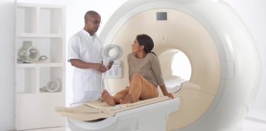
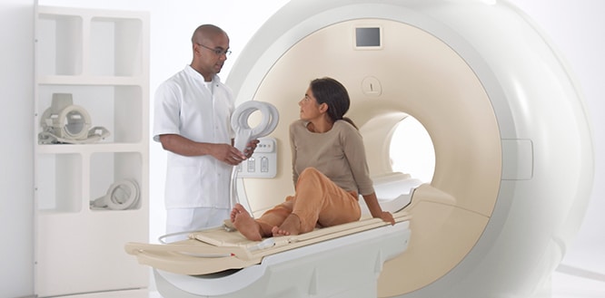
- The patient lies on a movable table that slides into the MRI device open on both sides.
- A specialist who monitors the patient from another room performs the procedure. The expert speaks with a microphone.
- Most people finish the procedure without any problem. People with claustrophobia may be given a sedative.
- The procedure is painless. People who have MRI do not feel the magnetic field or radio waves.
- Inside of the magnet creates repetitive sounds like pulling or hitting. Using ear plugs or listening to music can reduce noise.
- The shortest magnetic resonance scan takes 15 minutes. There are also MRI scans taking longer than one hour.
- During MRI, it is necessary to stand still to avoid blurring the images.
- During functional MRI, the patient may be asked to touch his thumb with other fingers or to answer some simple questions in order to check the parts of the brain.
- Those who complete the scanning may return to their life if they are not given sedatives.
How is medicated MRI taken?
- A contrast (dyeing) agent can be used for MRI scanning to obtain a more detailed image of the area. The substance called gadolinium is usually injected intravenously.
- It spreads through the circulatory system and provides a clearer view of the tissues.
- The duration of preparation and screening of the medicated magnetic resonance imaging is longer than that of a normal MRI.
- After the procedure, it is necessary to drink plenty of water to remove the contrast agent from the body.
Gadolinium rarely causes allergic reactions, but sometimes lead to side effects such as nausea, skin rash, headache. They are usually mild and short-term. However, a blood test may help determine how well the kidneys work in a kidney disease or whether it is safe to continue screening.
Patients with a history of allergic reactions, bleeding or clotting problems should always inform their doctors before any injection.
Things to do before an MRI
Cases before an MRI, the doctor should be informed:
- Health problems such as kidney or liver disease,
- A recent surgery,
- Allergy or asthma to foods or medicines,
- Pregnancy, or breastfeeding, especially if the contrast agent will be taken during the procedure,
- The presence of metal-based devices in the body.
Because MRI employs strong magnets, the presence of metals in the body can be dangerous when attracted to the magnet. Even if they are not affected by magnet, metal objects may disrupt the magnetic resonance image. Before an MRI, you must inform your doctor or technical specialist if your body has metal or electronic equipment.
These are metal-based devices that prevent access to MRI unless the MR device to be used is approved as safe:
- Metallic joint prostheses
- Artificial heart valves
- An implantable heart defibrillator
- Implanted drug infusion pumps
- Implanted nerve stimulators
- A pacemaker
- Metal clips
- Metal pins, screws, plates, stents or surgical staples
- Cochlear implants
- Type of a bullet, shrapnel or any piece of metal
- Intrauterine device
- If you have tattoos or permanent make-up, ask your doctor if they will affect your MRI. Be aware that some dark inks contain metal.
On examination day, the patient should wear large and comfortable clothes with no metal parts. During screening, the patient may be asked to undress and wear an apron. No preparation is required for scanning. The only exception is magnetic resonance cholangiopancreatography (MRCP), a special screening of the bile ducts. In this case, it is not allowed to eat or drink for 2-3 hours before the test.
It is not allowed to enter the MR room with mobile phone, coins, dentures, glasses, hearing aids, keys, jewelry, watches, hairpins, underwire bras, wigs, cosmetics containing metal parts.
Types of MRI
There are many types of MRI, but Diffusion Weighted MRI and Functional MRI (fMRI) are the main ones.
Diffusion Weighted MRI
This type of MRI identifies the character of tumors and diagnoses acute brain ischemia.
Functional MRI (fMRI)
It examines the anatomy of the brain and identifies which parts of the brain perform critical functions and which areas of the brain are active (consuming more oxygen) during various cognitive tasks. It can also assess damage due to head trauma or disorders such as Alzheimer’s disease. (6)
Perfusion Weighted MRI (PA – MRI)
Perfusion imaging is a dynamic examination using contrast agent, and helps diagnose and treat stroke, brain tumors and cerebral pathologies.
MRI Angiography
Magnetic resonance angiography (MRA) monitors blood vessels. It examines the areas of narrowing or expansion of blood vessels in the neck and brain. It can visualize the arteries that provide blood to the kidneys, as well as the vessels in the arms and legs.
Cardiovascular MRI
MRI focusing on the heart or blood vessels can assess:
- The size and functions of the chambers of the heart
- Thickness and movement of the walls of the heart
- Prevalence of damage caused by heart attack or heart disease
- Structural problems such as aortic aneurysms or dissections
- Inflammation or obstruction of blood vessels
MRI of the body
It is an MRI scanning from head to toe. It searches for cancers, infections and blockages in body. This scan takes approximately 45 minutes and starts from brain first. Then, neck, lungs, chest, liver, pancreas, kidney, adrenal gland, abdominal organs, bladder, prostate, testes, uterus are screened. Also, leg bones and muscles up to knee are examined in same session.
MRI of the breasts
MRI is used together with mammography to detect breast cancer, especially in women with dense breast tissue or high risk of disease. It diagnoses, treats and follows-up breast cancer and checks up the status of implanted breast implants.
Prostate MRI
3 Tesla Multiparametric MRI diagnoses early prostate cancer and determines accurate and precise location of the biopsy. It helps evaluate prostate cancer, abscess, enlargement, congenital abnormalities and complications after prostate surgery.
MRI of the Musculoskeletal System
This type of MRI helps to evaluate joint abnormalities, disc abnormalities in the spine, bone infections, bone and soft tissue tumors caused by traumatic or recurrent injuries such as torn cartilage or ligaments.
MRI for Brain and Spinal Cord
It is usually used for diagnosis of such conditions as:
- Aneurysms of cerebral vessels
- Eye and inner ear diseases
- Multiple sclerosis
- Spinal cord disorders
- Stroke
- Tumors
- Brain injury caused by trauma
Open MRI
Open MRI helps claustrophobic, obese and pediatric patients. Contrary to conventional MRI scanners, open MRI does not surround the entire body. Two or three sides are open. Open MRI makes the examination less stressful by providing a more comfortable, less restrictive environment and a lower noise level.
MRI Results
The radiologist analyzes the images in the scan, interprets the results and reports the findings to the doctor. How quickly the doctor will receive reports depends on the imaging center where the examination is done. The doctor will discuss important findings and the next steps with the patient.
Risks of MRI
- Noise: Loud sound, usually described as click and beep, with density up to 120 decibels on specific MR scanners, requires special ear protection.
- Nerve Stimulation: Rapidly changing areas in the MRI may cause a sense of twitching.
- Contrast agents: Patients with severe renal insufficiency requiring dialysis may carry a rare but serious risk of nephrogenic systemic fibrosis, which may be related to the use of certain gadolinium-containing substances.
- Pregnancy: Although there is no provennegative effect on the fetus, it is recommended to avoid MRI scans especially during the first trimester of pregnancy as the organs of the fetus are being formed.
- Claustrophobia (fear of confined space): People with mild claustrophobia may have difficulty in tolerating long scan times inside the device. Among the relaxation methods are being familiar with the device and the process, visualization techniques, sedatives and anesthesia. In addition, listening to music or watching videos, movies, closing, covering the eyes, and giving a panic button to the patient’s hand may help claustrophobic people during screening.
Does MRI have radiation?
MRI does not use ionizing radiation as used in X-ray and CT imaging; instead, it uses a strong magnetic field.