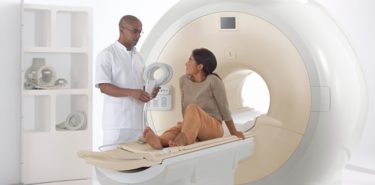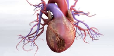
 Angiography (Angiogram) is an examination of blood vessels under X-rays using a kind of medical dye. It is performed by physicians in the angio laboratories. When the patient is lying on his back, a special sheath and then the catheter are moved up in the vein through the arteries in the groin or wrist. When the catheter reaches the heart, contrast material is injected and therefore imaging is possible under X-rays. This technique is most often performed for imaging of heart vessels. As a result of this procedure called coronary angiography, the obstructions in the heart vessels can definitively be diagnosed.
Table of Contents
What is Angiography?
Angiography is the process of imaging arteries under x-rays using a kind of medical dye (a contrast agent). Angiography (Angiogram) displays which region of the blood vessels is narrowed down. Although it is a compulsory method in people with coronary artery disease, it has risks such as allergy to contrast agent, heart rhythm disorders, renal failure due to contrast agent, stroke and heart attack.
However, these risks are unlikely to occur. If angiography is not performed due to these risks, the person is more likely to develop illness due to any damage to the heart.
To whom is angiography applied?
- Suspicion of coronary artery disease: Coronary angiogram is most commonly carried out for individuals at risk with a coronary artery disease after the required measurements and tests are done for a definitive diagnosis and to determine the stenosis of the blood vessels that feed the heart.
- Emergency situations: Urgent angiogram
- may be required for people who have a heart attack or who come back to life after an intervention in the hospital after a sudden cardiac arrest.
- Imaging of the veins of organs: Angiography is also used for imaging of the veins of the brain, neck, lung, kidney, arm and leg as well as the heart blood vessels.
Some of the diseases that require examination of the veins of these organs are:
- Vascular abnormalities: Aneurysm (vascular bubble formation), arteriovenous malformation (abnormal connection between artery and venous blood vessel) etc.
- Stroke: It sometimes occurs due to a decrease in the blood flow in the brain due to blockages in the carotid artery and branches.
- Peripheral artery disease: Congestion of arm or leg veins
- Renal artery stenosis: Contraction of the blood vessels that feed the kidney
- Dissection: Separation due to various reasons in the layers forming the inner wall of large blood vessels such as aorta
- Pulmonary embolism: Clot in the blood vessels that feed the lung
- Congenital heart and vascular abnormalities, and diseases which commonly involve blood vessels
What does angiogram do?
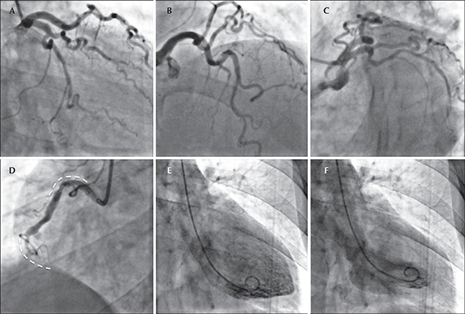
After the end of the coronary angiogram, digitally recorded images are evaluated by the physician who performs the procedure, and a report is written as to whether the blood vessels supplying the heart are narrowing down, which blood vessels narrow and the percentage of the narrowing. Treatment is administered according to the outcome of the report.
These treatments include balloon angioplasty, stenting, bypass surgery or blood thinners. The decision on which treatment to apply is taken by the doctor according to the degree of stenosis, how many vessels are affected, and the characteristics of the patient.
Why is angiogram performed?
- When complaints of the heart are repeated despite adequate medical treatment
- If ECG, echocardiography, or effort ECG test is negative when performed on people with chest pain (angina) that do not suggest coronary artery disease
- If heart failure develops after a heart attack
- When people who are at risk for coronary artery disease have chest pain (angina)
- If people who have previously been intervened into their heart vessels by balloon angioplasty, stenting, or bypass procedure have pain or a heart attack again
- If a patient with sudden cardiac arrest survives as a result of intervention
- In order to diagnose an emergency after a heart attack
- If life-threatening heart rhythm disorder is detected
- If there are symptoms and signs suggestive of ischemic heart disease in people with heart valve disease
How long does angiography take?
Coronary angiogram takes approximately 15-20 minutes. However, due to various disorders in the vascular structure of the patient, the procedure time may be prolonged. In addition, angiography can be prolonged because of the need to intervene in the case of any problem that occurs during angiogram.
After angiography, pressure should be applied to the catheter insertion point for approximately 15 minutes in order to avoid bleeding while the catheter is removed. If the application area is in the groin, you will need to lie on your back for about 6 hours since a sandbag will be placed for bleeding control.
If the area of application is the veins on the wrist, there is no need to place a sandbag and a long stay is not required.
Things to do before angiography
- From 12.00 a.m. on, do not eat or have breakfast on the day of the operation.
- Bring your medications to the hospital. You can take your mandatory medications after asking your doctor.
- You should drink plenty of water to prevent kidney damage after angiography.
- Do not have any items such as rings, earrings or glasses on you.
- You should have a companion who can stay with you after angiography and help you go home.
- Angiography can be performed from the arteries of the arms or vessels of the groin. Shave both groin areas in case groin veins are used.
- Read the consent form given by the doctor. Sign if you agree.
Preparing for an angiografy
- Before angiogram is performed, your doctor will ask you about your medical condition, additional diseases, medications you use and if any, your allergies.
- Complete blood count (hemogram), clotting values, liver and kidney function tests, values of important minerals in the body, such as sodium, potassium, and tests to measure heart enzymes as a sign of heart attack are done.
- Pulse examination is performed. In this way, it is decided whether the angiography will be performed in the groin or wrist artery.
- Tell your doctor if you are taking blood thinners (such as warfarin). If necessary, the medication you use will be discontinued and other medicines that give the same outcome will be recommended.
- Have all test results with you until angiography is done.
- If angiogram, bypass, stent has been performed before, take these test results with you
How is angiography performed?
- It is performed by doctors specialized in this subject in a specially equipped laboratory.
- The patient is awake during the procedure. If necessary, sedatives may be given.
- The patient lies on his back on the angiography table.
- He/she is connected to machines that show the heart rhythm, blood pressure and oxygen levels in the blood.
- The procedure can be performed from the groin or wrist. That area is cleaned with an iodized antiseptic solution and sterilized.
- The patient is covered with sterile drapes.
- The area where the catheter will enter is anesthetized.
- The arteries in the groin or wrist are entered by means of a special needle and a sheath is inserted at the entrance.
- The special soft thin tube called catheter is advanced through the sheath to the main veins of the heart. Progression within the veins does not cause pain and is not felt by the patient.
- The contrast material (a kind of dye) is delivered to the heart via the catheter.
- Images of the blood vessels are taken by X-rays while the heart is working. These images are recorded digitally. Doctors can then watch them in order to write the report.
- Coronary angiography is painless. Only a needle prick is felt when the area to be treated is anesthetized.

Risks and complications of angiography
Angiography is a safe process. Rarely, undesirable conditions may occur during or after angiography. However, if angiography is required and if it is not done, it is more likely to cause problems. Some of the problems that may occur due to angiography are:
- Allergy to contrast agent
- Pain, swelling, bruising at the catheter insertion point in the groin or wrist
- Infection at catheter insertion point
- Renal failure due to contrast agent
- Heart rhythm disorders
- Need of a temporary pacemaker
- Stroke
- Heart attack
- Bleeding due to vessel damage
- Junction between artery and vein that may occur during sheath placement during angiography (arteriovenous fistula)
- Death: Angio-related death is very rare.
Problems after angiography
In the groin or wrist region where the catheter is placed
- Bruising and pain
- Sensitivity and discomfort
- A slight swelling may occur.
During pain felt while pulling the catheter out from the groin or wrist area, the so-called vagal reaction may occur. In this case, nausea, cold sweating, blood pressure and low pulse are seen.
Post-angiography edema and fatigue
Swelling of the arms and legs or edema may occur on the side where angiography is performed. This is not a normal condition and should be told to the doctor. It is a condition caused by the damage of the veins in the arms and legs.
In case of renal failure due to contrast agent used in angiography, widespread swelling and edema may occur in the body due to the inability to remove fluids from the body. Excessive bleeding due to angiography may cause fatigue.
What to do after angiography?
- When the angiography is over, if there is no serious condition, you will be taken to your room in the hospital. If serious angio-related problems develop, you may need to be followed up in the intensive care unit.
- Once you are taken to your room, you are connected to machines that show the heart rhythm, blood pressure and blood oxygen levels.
- After angiography, catheter is removed from the wrist or groin. In order to prevent bleeding in that area, pressure is applied to the area by the personnel concerned. You may feel pain during this time.
- After withdrawing the catheter, sandbags are placed to prevent bleeding. You should lie in the supine position as long as there is a sandbag. After about 4-6 hours, bleeding is checked and the sandbag is removed.
- Do not get out of bed before this process is over.
- Once the sandbag has been lifted, rise slowly as you may experience dizziness when you first get out of bed.
- In case of coughing or sneezing, apply pressure manually to the treated area.
- Drink plenty of water for easy removal of the contrast agent, which is harmful to the kidneys.
- You may be discharged on the same day after angiography, but sometimes there may be situations where you need to stay in the hospital for additional tests and treatments.
- Ask your doctor what to do and not to do after discharge.
- Avoid constipation and severe strain.
- Do not lift heavy, do not drive for 2-3 days.
- Protect the operation area from impacts.
- Follow the treatment initiated by your doctor.
WARNING:
If you experience one or more of the following symptoms after angiography, consult your doctor or emergency room immediately:
- Severe pain that does not go away with painkillers
- Widespread flushing, swelling, heat increase on skin at the catheter insertion point
- Unstoppable bleeding at the catheter insertion point
- Different image of the treated arm or leg comparing to the arm and leg on the other side
- Inability to urinate, decrease in urine output
- Having a tight, tense swelling at the catheter insertion point
- Shortness of breath and chest pain
References: 1- Preparing for an angiogram, 2- Angiography, 3- Coronary Angiography