Radiology is a branch of medicine which uses various imaging technologies in the diagnosis and the treatment of diseases. Radiological examinations are carried out in almost every field of healthcare industry. Radiology uses different imaging methods such as X-ray, magnetic resonance imaging (MRI), computed tomography (CT) and PET scan to see internal body structures. It is divided into two branches named diagnostic radiology and interventional radiology. Radiology is a vital component of most medical decisions affecting patients, and radiologists can work with all clinical specialties.
Table of Contents
What is radiology?
Radiology is a medical specialty that interprets the internal body structures to diagnose diseases. It creates internal body structure images using radiation, ultrasonic sound waves or extremely strong magnetic fields for the diagnosis and the treatment of diseases. A specialist examining the comprehensive diagnostic tests is called a radiologist. A radiologist is specialized in the diagnosis of diseases and injuries.
Diagnostic radiologists are the last step in diagnosis process. They examine the images which may be beneficial to evaluate and support a treatment. A medical imaging specialist who can use and manage the technical equipment to generate images is called a radiologic technologist. While the department in which radiological examinations are performed is often called radiology department, it may also be named radiography department or medical imaging department. (1)
What diseases does radiology treat?
According to a patient’s condition, radiologists may cooperate with every medical specialist. The main focus of diagnostic radiology is to define and monitor diseases, abnormalities of skeletal and soft tissue, and trauma. Radiology is essential in the diagnosis of many diseases, especially cancer. (2) It is also used in:
- Diagnosis of bone and lung diseases
- Examination of foreign material
- Injuries and emergency medicine
- Breast diseases
- Diagnosis of osteoporosis
- Diagnosis and treatment of cardiovascular diseases
- Diagnosis of internal organ diseases
- Follow-up of pregnancy
- Diagnosis of musculoskeletal diseases
- In the nervous system examinations such as spine, spinal cord, brain, head, neck, waist, neuroradiology
- Imaging of soft tissue lesions in abdomen and breasts
- Examination of gynecological and pediatric diseases.
Nuclear medicine practices including bone scan, thyroid scan and thallium stress test are included in the field of radiology.
Thanks to technological developments that enable electronic storage of radiological images, outbreaks can be detected faster and more accurately.
Branches of radiology
Diagnostic radiology
Diagnostic radiology may be used to: (3)
- Define the cause of symptoms
- How well the body responds to an administered treatment
- Diagnose various diseases such as breast cancer, colon cancer and cardiac diseases early.
In diagnostic radiology, the images of internal body structures are obtained and diagnostic radiologists interpret these images.
Most commonly used diagnostic radiology tests:
- Computed tomography (CT) including CT angiography
- Fluoroscopy, which includes the upper GI (gastrointestinal system) series and barium enema
- Magnetic resonance imaging (MRI) and magnetic resonance angiography (MRA)
- Mammography
- Nuclear medicine examinations including bone scan, thyroid scan and thallium stress test
- X-ray including chest X-ray
- Positron emission tomography, also known as PET imaging or PET scan
- Ultrasound
Interventional and therapeutic radiology
Interventional radiology provides imaging guidance for minimally invasive procedures in the treatments of the patients who do not undergo an open surgery. Imaging methods such as CT, ultrasound, MRI and fluoroscopy are used to help guide medical procedures. .
Imaging guides doctors while they insert catheters, wires and other small instruments and devices into the body. Therefore, the procedure can be performed with no incision or a small incision.
Examples of interventional radiology procedures:
- Angiography (vascular imaging) or angioplasty (vasodilation) and stent placement.
- Embolization to control a bleeding.
- Cancer treatments: Chemoembolization (embolization with microspheres filled with chemotherapy drugs) or tumor embolization with Y-90 radioembolization (Yitrium-90 microsphere)
- Tumor ablation treatments: Radiofrequency ablation (evaporation of a tumor using heat), cryoablation (freezing a tumor) or microwave ablation (evaporation of a tumor using heat)
- Vertebral and spinal fracture treatments: Vertebroplasty and kyphoplasty
- Needle biopsies: In lungs, thyroid gland or different organs
- Breast biopsy: Stereotactic or ultrasound techniques
- Uterine artery embolization
- Feeding tube insertion
- Venous access catheter placements such as port catheter and PICC catheter
Diseases for which Interventional Radiology is used
Interventional radiology is often used in the treatment of cancer or tumors, clogging in the arteries and veins, uterine fibroids, back pain, liver and kidney problems. This type of treatment has fewer risks than traditional surgery, causes less discomfort with a shorter recovery period. (4)
Diagnostic Equipment in Radiology
X-Ray
X-ray, which is generated by X-rays, is used to examine bones, chest and abdomen and foreign bodies. A fluoroscopy may be performed to obtain a dynamic image of the digestive system. Fluoroscopy may be performed with a contrast agent to perform an angiogram showing the blood vessels. (5)
Computed Tomography (CT)
A CT generates cross-sectional images (slices) of a person’s body with multiple X-rays directed to an intended area. It is called “computed tomography” as it produces the images based on the calculations of a computer. It can examine the functions of the body and organs. It uses radioactive tracers circulating in the body while generating the images. (6)
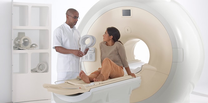
Magnetic Resonance Imaging (MRI)
It produces high-quality images of body structures using magnetic fields and radio waves with the help of a computer program. This technique that generates images by using of the potential energy stored in the body’s hydrogen atoms is called Magnetic Resonance Imaging. It does not use radiation. (7)
Ultrasound
It uses sound waves to generate dynamic images on a monitor. It is the most cost-effective and harmless imaging method. Ultrasound probes (transducers) use acoustic energy above the threshold level of hearing of humans to generate images. As ionizing radiation is not used in ultrasound, it is especially preferred to obtain images of children and pregnant women. (8)
Mammogram
Mammogram is an imaging method using X-ray specifically enhanced and positioned for breast tissues. Mammogram is the most ideal test for the early diagnosis of breast cancer. (9)
Fluoroscopy
It is the technique of obtaining real-time and dynamic images of the body with the help of X-rays for procedures such as stent placements for narrowed vessels or drainage catheters, or the imaging of the gastrointestinal tract. The radiation dose is higher than traditional radiography. (10)
Nuclear medicine
A radioactive substance with a short half-life is given to a patient to generate nuclear medicine images. Then, the radiation emitted from the patient is recorded and processed on a computer.
Who is a Radiologist?
What does a radiologist do?
Radiologists are doctors who can observe and interpret medical images for the diagnosis and treatment of diseases or injuries. They have advanced knowledge in anatomy and pathology fields. Their branches are divided as “diagnostic” or “interventional”, although many of them practice both.
Diagnostic radiologists can specialize in different sub-branches of radiology through extensive clinical practice and related studies:
- Mammogram
- Cardiovascular radiology (heart and the circulatory system)
- Chest radiology (heart and lungs)
- Emergency radiology
- Gastrointestinal radiology (stomach, intestine and abdomen)
- Genitourinary radiology (the reproductive and urinary systems)
- Head and neck radiology
- Musculoskeletal radiology (muscles and bones)
- Neuroradiology (brain and the nervous system, head, neck and spine)
- Pediatric radiology
What is the role of a radiologist?
- Interprets the findings of an imaging examination and writes diagnostic reports to assist doctors and healthcare professionals.
- Helps other doctors and healthcare professionals choose the appropriate imaging tests for their patients. Works as a part of the clinical team to balance the risks and benefits of imaging and prevent unnecessary radiation exposure.
- Is often asked to perform image-guided procedures such as biopsy, drainage, and targeted injections. Some radiologists may also perform more advanced image-guided procedures such as vascular stenting and aneurysm coiling
How to become a radiologist?
A person who wants to become a radiologist applies to radiology postgraduate education after completing his medical school education. After passing the exams in this 4-year education, if you are considering a sub-specialty, it is necessary to apply for a 1 or 2-year scholarship program. Although the practice of each state is different, often the doctor must obtain a license in the state where he or she is located.