 Breast cancer is a malignant tumor that is usually seen in the milk ducts and glands within the breast tissue. It mostly impacts women and is the most common type of cancer worldwide after lung cancer and cervical cancer. Statistics show that one in eight women will develop breast cancer at some point in their lifetime. Its main symptoms are lump or swelling in the breast or armpit, breast deformity and nipple discharge. Today, if diagnosed early, breast cancer can be cured. Therefore, women who are older than 50 years old and have a family history of breast cancer should regularly visit their physicians and undergo screening by mammography.
Table of Contents
What is breast cancer?
Breast cancer is a tumor that is formed when cells in the breast grow out of control and has potential to spread to other organs. As with other types of cancer, breast cancer is a tumor formed as a result of abnormal growth and proliferation of cells due to the faults in DNA. Tumors can either be a benign tumor or a malignant tumor. Benign tumors are not cancerous; they have cells that are normal and grow slowly and do not invade the surrounding tissues.
Malignant tumors are defined as cancer; they proliferate rapidly and spread to other organs (metastasis) when left untreated, eventually leading to death.
Breast cancer symptoms
- Painless, firm mass on the breast or armpit
- Visible abnormal change on the size or shape of the breast,
- Orange peel appearance on the breast skin,
- Development of wound, redness, bruise, edema or swelling on the breast skin
- Bloody or transparent discharge from the nipple
- Nipple inversion or deformity
- Changes of peeling and crusting on nipple skin
Causes and risk factors of breast cancer
- As a primary risk factor “being a woman”
- Being over 50 years old
- Diagnosis of breast cancer in a first-degree relative such as mother or sister
- No history of birth or breastfeeding
- Having the first birth after the age of 30
- Early onset of menstruation (for example before the age of 12)
- Entering the menopause after the age of 55 (late menopause)
- Receiving hormone therapy after menopause
- Using birth control pills for a long time before the first birth
- Gaining excess weight
- Alcohol and tobacco use
- Radiotherapy exposed at a young age (for example before the age of 15)
- Previous history of cancer in one of the breast
- Low fat ratio of the breast tissue
- Carrying breast cancer gene (BRCA)
Early diagnosis of breast cancer
Early diagnosis in breast cancer both greatly facilitates the treatment and increases the rate of success. The goal of early diagnosis is to find the cancer before it causes symptoms. The most important factors determining the outcome of treatment in breast cancer is the size and spread of the tumor.
Tests which allow early diagnosis of breast cancer save thousands of lives every year. First of all, it should be known that 8 in 10 breast masses are benign, meaning they are not cancer. However, if you notice a palpable and unusual formation or mass in the breast, you must seek help from a specialist.
Best steps for diagnosis of breast cancer
Mammography for breast cancer
Women over the age of 40 are recommended to take annual mammographies. Mammography is the X-ray of breast. All procedure takes approximately 20 minutes. As it may not always be adequate for cancer diagnosis, different methods of screening such as ultrasound or MRI (magnetic resonance imaging) may also be recommended in women with high risk of breast cancer.
Clinical breast exam
Clinical breast exam (CBE) by a specialist preferably every 3 years at twenties and thirties and every year after the age of 40 is highly important for early diagnosis. Also, if you do not know, you can learn how to perform breast self-exam during these exams.
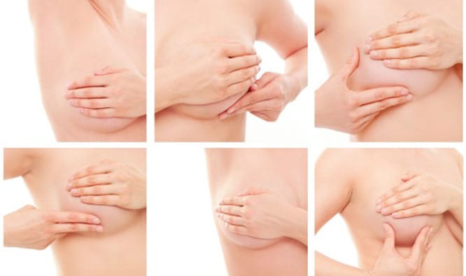
How to perform breast self-examination?
Knowing the symptoms of breast cancer is very important to prevent the advancement of breast cancer. Therefore, an individual should know their breast structure and risk factors. To notice the symptoms of breast cancer, every woman should perform monthly breast self-exams after the age of 20. Thereby, they can get to know their breast structure and notice the newly developed masses early.
Breast self-exam should be performed 5-7 days after the end of menstruation when the effects of the hormones are at minimum; and women who are not menstruating should perform this exam any day of the month they wish, however, it should be performed on the same day of every month.
Steps to Breast Self-Examination
In front of the mirror
- Get in front of the mirror bare-chested and with your arms hanging freely on the sides, check if there is any difference in the size, shape, texture or skin of your breasts.
- After this, put your hands on your hips and press firmly. Observe the outer parts of your breast by turning to both sides.
- Rotate your shoulders and elbows to the front and bend over to contract your breast muscles; your breasts will fall frontward. In the meantime, examine for changes of shape and outer line in your breast.
- Fold and firmly grip your hands on the back of your hands and stretch your elbows backwards. Check the outer surfaces and especially undersides of your breasts by turning to both sides. Lift your breasts with your hands to see this region, if necessary.
- To observe whether there is a discharge coming from the nipple, place your index finger and thumb on the area surrounding the nipple, and pull the nipple towards the end. Repeat this for your other breast.
During bath
- To feel the changes in the breast easier, lubricate your hands using soap. Raise your one arm upwards. Put your 2nd, 3rd and 4th fingers of your other hand together for examination, and gently press your skin by using the inner surfaces of your finger, and check all your breast and armpit area. Thereby, you can notice the densities on the upper outer breast quadrant easier.
- Starting from the armpit, draw an imaginary and large circle surrounding the breast. Examine all area by gradually shrinking the circles towards the nipple. Later, move your finger up and down starting from armpit again. Continue movements by shifting towards the inner part and be sure that you touch the lower margin of the breast.
- Make a star-like motion with your finger from the outer margin of the breast towards the nipple, and check all breast tissue. And check the armpit again.
In supine position
- Lay down and place a small pillow or a folded towel under your right shoulder. Place your right hand to the back of your head. Again with your fingers put together, place your left hand on the upper part of your right breast. For an easier experience, you can use body lotion or oil.
- Make small clockwise circles and examine the whole surface back to the starting point. When the circle is completed, repeat this same circular motion this time around the nipple. Continue like this until all breast surface is screening. Be sure that you also examine the more upper parts towards the armpit.
- Place your fingers directly on your nipple in straight position, and check for any change around it. Gently press the nipple in. Be sure that it easily moves. Later, repeat the same steps for your other breast.
- In case of high risk: Under the regular follow-up of their physicians, women at high risk for breast cancer may need to start taking mammographies earlier, undergo additional workup or get examined more frequently.
Suspicious breast lumps
If there is any reason suggesting that you may have breast cancer, your physician may recommend some of following tests after complete examination; however, none of these individual tests are used for one individual. When choosing a diagnostic test, your physician should also take your symptoms, age, medical history and results of your previous tests into account.
Imaging tests used to diagnose breast cancer
Diagnostic Mammography
While mammographies are mostly used for screening, they are also used in case of cancer suspicion and this is called diagnostic mammography.
Breast ultrasound
Ultrasound uses sound waves to take images of a body region. Ultrasound became a good method to be used together with mammography. Also, using an ultrasound, it is possible to distinguish a mass from a usually non-cancerous fluid-filled cyst.
Ductography
While it is a special X-ray technique, it sometimes help determining the cause of nipple discharge.
MRI (magnetic resonance imaging)
An MRI uses does not use X-rays but magnetic fields to take detailed body images. Also, it can be used together with mammography for woman at high risk for breast cancer.
Biopsy
If other tests indicate cancer, the definitive way of diagnosis is biopsy. During this procedure, cells are taken from the area of interest and examined under microscope. There are many types of biopsies:
- Fine needle aspiration biopsy: A fine needle is used to extract a small cell sample. If the biopsy does not provide a definitive result or your physician is still not sure, a second biopsy or a different type of biopsy may be necessary.
- Stereotactic core needle biopsy: A thicker needle is used to extract a larger or several tissue samples. It does not require hospitalization; the area is anesthetized using local anesthesia.
- Surgical biopsy: The procedure of removal of the lump partly or completely. This procedure is usually performed using local anesthesia without hospitalization.
- Biopsy laboratory tests: Tissues taken by biopsy are examined in laboratory, and differential diagnosis for benign or malignant masses is made. If there is no cancer, no additional treatment is necessary. If it is cancer, it gives us information about the type and growth rate of the cancer, and helps the physician mange the treatment process.
Diagnostic tests for metastatic breast cancer
- Pulmonary X-ray: This test is performed to see if the cancer is spread to lungs.
- Bone scintigraphy: Being performed to see if the cancer is spread to bones, this workup exposes the patient to a small dose of radiation.
- CT (computerized tomography): CT is a special type of X-ray which takes many images from various angles. This workup can be used to determine whether the cancer is spread to the liver or other organs. Also, it can be used as a guide to place the biopsy needle into the suspected area.
- MRI (magnetic resonance imaging): Being a little bit more uncomfortable compared to CT as it takes longer to take and is taken in a narrower area, this imaging method allows us to observe the spread of the cancer to the brain and spinal cord.
- PET (positron emission tomography) workup: This workup is used if the spread is known but it is not known where. Cancer cells are observed using a type of sugar containing a radioactive atom and a special camera. Because cancer cells absorb abundant amounts of sugar. Also, it can be used for cancer control before lymphatic glands are removed.