Computed Tomography (CT) scan is a medical imaging technique using X-rays and a computer to give us detailed images of the internal parts of the body. Tomography helps view almost all parts of the body. It is useful to diagnose diseases or injuries, as well as to plan surgical interventions or treatments. It can save lives in emergencies. Since CT scan uses radiation, risks and benefits should be taken into consideration.
Table of Contents
What is CT scan?
Computed Tomography, is forms cross-sectional images of the bones, blood vessels and soft tissues of the body by computing a series of X-ray images taken from different angles around the body. It describes normal and abnormal structures in the body or assists medical procedures. CT scan images provide more detailed information than plain X-rays.
A contrast material containing iodine is sometimes used for screening. It is painless, and is often performed in an outpatient setting. (1)
How does a CT scanner work?
A large x-ray source and an x-ray detector with a circular aperture in the center rotates around the body. Modern scanners usually contain an x-ray detector with 4 to 64 or more sensors that record x-rays passing through the body. The data coming from the sensors makes a series of X-ray measurements taken from multiple angles in the body. Measurements are not displayed directly, they are sent to a computer.
The computer converts x-ray measurements into images reminiscent of 2-dimensional slices (cross-sections) of the body. Tomo means “slice” in Greek. This recorded image is called a tomogram. The process is repeated to produce some sections. The computer can also make 3D images from the recorded images. (2)
What is CT scan used for?
Tomography helps diagnose damages and injuries in the body, guide further examinations or treatments, and control the effects of treatments.(3)
The CT scan is particularly used to:
- diagnose muscle and bone disorders such as bone tumors and fractures,
- identify problems related to bone damage, blood flow, stroke and cancer,
- determine the location of the tumor, infection or blood clot,
- guide procedures such as surgery, biopsy or radiation therapy
- identify and monitor diseases and conditions such as cancer, heart disease, lung nodules and liver masses,
- follow-up the effects of some treatments such as cancer treatment,
- detect internal injuries and internal bleeding,
- guide the radiologist in performing specific procedures such as a biopsy, extraction of intra-bodily fluids for various tests, or evacuating deep abscesses,
- determine the location, size and shape of a tumor.
What can CT scans detect?
- Brain problems: Brain tomography can detect traumatic injuries (such as blood clots or skull fractures), tumors, and infections.
- Spine problems: The bone structure of the spine, the intervertebral disc between the vertebrae and the anatomy of the spinal cord can be accurately defined.
- Osteoporosis: CT scans can be used to accurately measure bone density in assessing osteoporosis.
- Intra-abdominal diseases: Abdominal CT scans are used to confirm the presence or absence of changes in the body caused by tumors, infection, abnormal anatomy or trauma. They help define the anatomy of the body, including visualization of the liver, gallbladder, pancreas, spleen, aorta, kidneys, uterus and ovaries.
- Heart diseases: CT scan of the heart is useful when various types of heart disease or abnormalities are suspected.
- Intracranial traumas: CT scan can be beneficial for head imaging to detect traumatic injuries, tumors, paralyzing clots, bleeding, tumors and infections.
- Pulmonary diseases: It can monitor the lungs in order to detect other conditions such as tumors, clots, excess fluids and emphysema or pneumonia.
- Bone diseases: CT scanning is particularly helpful when imaging complex bone fractures, severely worn joints, or bone tumors.
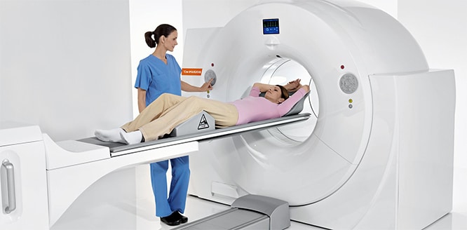
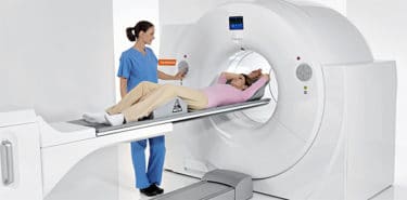
How is CT scan done?
- You may be asked not to eat or drink a few hours before CT.
- Before scanning, a special dye called contrast can be given to the body to help improve the quality of the images.
- During the CT scan, some or all of clothes are taken off and a hospital gown is worn.
- Since they affect the screening results, metal objects such as belts, jewelry, dentures, clasps, piercings, glasses are removed.
- You lie prone or supine on a table that slides or stays fixed to the scanner. In addition, belts or pillows can be used to maintain the position.
- The radiologist leaves the scanning room and enters the control room. There is a system where the specialist can see and communicate with the patient.
- As the table slowly enters the scanner, the X-ray device rotates around the patient. Numerous images of thin sections of the body come out at each rotation.
- Since movements may cause blurry images, it is very important to stand still when capturing CT images.
- In addition to breathing or exhaling during the examination, you may be asked to hold the breath briefly to avoid chest movement.
- Sounds like clicks or buzzing may be heard during scanning. Also, the table may do millimetric movements.
- The doctor may recommend a sedative to keep young children calm and still during CT scans.
How long does a CT scan take?
The whole process usually takes a few minutes to 20 minutes. It can be extended up to 1 hour. Persons who undergo a CT scan can return to their daily routine after the scan. If contrast is used for screening, you may be advised to wait in hospital for about an hour.
How do you prepare for a CT scan?
- Inform your doctor and hospital if you have any allergies or kidney problems, or if you are taking medications for diabetes.
- Tell your doctor if you are pregnant. CT scans are not recommended unless it is an emergency for pregnant women because X-rays are likely to cause harm to the baby, albeit low.
- Do not wear zippered or jewelery clothes on day of screening, prefer comfortable clothes.
- Unlike MR scans, the scanner does not cover the whole body, so claustrophobia is not a concern. However, if you are worried, doctors may prescribe sedatives.
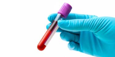
How is a CT scan with contrast done?
Dense substances such as bones are easily visualized in CT scans. However, soft tissues may appear pale on x-ray images. For a clear image, a special paint, sometimes called contrast agent, can be used. The contrast agent blocks x-rays, emphasizing blood vessels, organs or other structures and makes them appear white in the scan. Contrast agents are usually made of iodine or barium sulfate. (4)
They are given to the body in different ways depending on the tissue and organ to be screened:
- Intravenous (IV) contrast: Drugs are injected directly into the vein. It helps blood vessels, urinary tract, liver or gall bladder to stand out in the image. During the injection, warmth or a metallic taste may be felt in the mouth.
- Oral contrast: You may be asked to drink a liquid containing contrast agent for screening the esophagus or digestive system. The drink may taste bad.
- Enema: If the intestines are scanned, the contrast agent can be put in the rectum. This may cause bloating and discomfort.
After the CT scan, it is recommended to drink plenty of fluids to get rid of the contrast agent.
Different types of CT scans
Brain tomography (Cranial CT) scan
Cranial CT scan is also known as head scan, skull scan and sinus scan. It uses a special x-ray machine to assess head injuries, severe headaches, dizziness and other aneurysms, bleeding, stroke and brain tumor symptoms. It also helps to assess the face, sinuses and skull, or to plan radiation therapy for brain cancer. (5)
Spine CT scan
Spine CT helps to evaluate the structures in and around the vertebrae in the neck (cervical), back (thoracic) or lumbar region. It is an imaging method used to diagnose spinal cord’s injury. In an emergency, it may indicate internal injuries and bleeding. (6)
Chest CT
The special x-ray machine of computed chest tomography examines abnormalities found in other imaging examinations and investigates the cause of unexplained cough, shortness of breath, chest pain, fever and other chest symptoms. Since it can detect even very small nodules in the lung, it is especially helpful in diagnosing lung cancer at its earliest and curable stage. (7)
Abdomen and Pelvis CT scan
Abdominal and pelvic tomography is a diagnostic imaging test identifying diseases in the small intestine, colon and other internal organs, and often determining the cause of unexplained pain.
CT Angiography
Computed tomography angiography is used to diagnose and evaluate conditions such as blood vessel disease, aneurysms or obstructions. Contrast material is injected into the blood vessels. CT angiography helps visualize the brain, neck and heart vessels, and large vessels in the rib cage and in the abdomen, heart vessels, as well as arm and leg vessels.
How long does it take to get CT scan results?
Scan results are usually not available immediately. Information in the scan needs to be processed by the computer and analyzed by a radiologist. After analysis of the images, the radiologist writes a report and sends the results to the doctor who referred the patient to the scan. Then, the doctor shares the results with the patient.
CT scan results are considered normal, if the radiologist does not see any tumors, blood clots, fractures or other abnormalities in the images. If any abnormality is detected during a CT scan, the patient may need other tests or treatments depending on the type of abnormality found. This process can take from some days to a few weeks.
Risks of CT Scan
CT scans are fast, painless and usually safe. Nevertheless,
- Allergic reaction: In some patients, the contrast agent may cause allergic reactions or, in rare cases, temporary renal failure. Hypersensitivity reaction to contrast agents may range from rush to anaphylactic shock.Risk factors for hypersensitivity to intravenous contrast are allergic reactions, asthma, or food allergies. Patients with severe conditions such as renal failure/insufficiency, multiple myeloma, congestive heart failure, aortic stenosis should not take contrast agent in the vessel.
- Renal toxicity: Renal toxicity, which may result in renal insufficiency, is a very rare complication of intravenous contrast agent used in CT scans. This reaction is more common in people with diabetes, exposed to dehydration, or in patients with previous impaired renal function.
- Rare complications: Complications such as bleeding, infection, shock, and death as a result of the procedure may rarely occur due to procedural interventions.
- Ionizing radiation has the potential to cause biological effects in living tissues. This risk may increase with the number of CT scans that a person has taken throughout his or her life. The amount of radiation exposed during a CT scan varies depending on how much of the body is being scanned.
Exposure to radiation during CT scans may slightly increase the likelihood of developing cancer after several years, but this risk is considered very low (less than 1 in 2,000).
CT scans during pregnancy
If there is no abdominal or pelvic imaging during pregnancy, there is no risk for the baby. If imaging of the abdomen and pelvis is required, non-radiation tests such as MRI or ultrasound are preferred. CT can be tolerated in case of an emergency. Nevertheless, CT carries a potential risk to the fetus in the first trimester of pregnancy.
Is a CT scan safe for a child?
Children are more susceptible to ionizing radiation, therefore cancer development is a higher risk in children than in adults. So, it is important to ensure that the scanner is adjusted for children during tomography.