Ultrasound is a medical imaging technique to examine the body and organs. It is very common in diagnosis, treatments and biopsies. There are two kinds of ultrasound: diagnostic and therapeutic. Diagnostic ultrasound, which is often applied to the skin, produces images through high-frequency sound waves produced by a transducer (probe). Nevertheless, for a better view, a diagnostic ultrasound probe can also be inserted into the body through either gastrointestinal tract, vagina or blood vessels. This type of ultrasound also works using sound waves, but does not produce images. It is used to move and push tissues, warm them up, dissolve blood clots, or deliver medication to specific parts of the body. Standard ultrasound has no harmful effect.
Table of Contents
What is Ultrasound (USG)?
Ultrasonography, sonography and ultrasound scanning, produce real-time images of organs and other structures in the body using high-frequency sound waves. The frequency of the sound waves is greater than 20kHZ and above the threshold that human ear can hear. Sound waves are produced by a transducer that emits ultrasound waves and can detect echoes. This USG probe sends high-frequency sound waves to the body. (1)
How does ultrasound work?
When a sound wave hits a dense object such as organs or bones in the body, it is reflected back or creates an echo. The resulting echoes produce electrical signals by reflecting back to the probe, and then send them to the scanner.
Echoes and their distances measured by a computer produce a moving image monitoring different densities of tissue, fluid and air in the body. One or more motion pictures are captured as still images. USG images can be two-dimensional (2D), three-dimensional (3D) or 4D (motion 3D). (2)
What does ultrasound do?
USG examinations help diagnose various diseases and assess the damage to the organs caused by the disease. It is a screening that is frequently used for the investigation of symptoms such as pain, swelling and infection, and examination of most internal organs of the body.
What an ultrasound can detect?
Circulatory problems
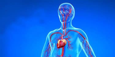
Doppler ultrasound, a special ultrasound technique: (3)
- Measures the direction and velocity of blood cells during vascular movement,
- Examines blood flow or blood pressure in a vein.
- Determine the velocity of blood flow and blockages.
- Diagnoses various heart diseases including heart valve problems and congestive heart failure, and assesses the damage after a heart attack.
Ultrasound of the heart is called “echocardiography” or “echo”. ECO gives images of the cardiovascular system and helps to measure cardiac tissue movements. In addition, it is used for:
- Examining the walls of blood vessels,
- Checking for DVT (Deep Vein Thrombosis) or aneurysm,
- Checking fetal heart and heartbeat,
- Investigating plaque accumulation and clots,
- Examining obstructions or narrowing of arteries,
- Checking tumors and congenital vascular injuries,
- Examining the heart and blood vessels, including the aorta and its major branches,
- Monitoring the decrease, absence or increase of blood flow in various organs such as testis or ovary. Information about the velocity and volume of blood flow obtained from the Doppler ultrasound image can determine whether the patient is a suitable candidate for a procedure such as angioplasty (dilation of arteries).
Ultrasound in emergency medicine
It is used to assess conditions such as:
- Traumatic wounds,
- Pericardial tamponade,
- Accumulation of fluid around the heart,
- Hemoperitoneum or blood leakage in abdominal area.
Abdominal ultrasound
Gastroenterologists use USG for getting images of spleen, kidney, biliary tract, gallbladder, liver, aorta, lower vena cava, pancreas and other solid organs within the abdomen. They benefit from ultrasound to diagnose gallbladder inflammation, known as suspected gallbladder stones or cholecystitis. USG can detect swelling or infection in appendix, which is a sign of appendicitis. (4)
Newborn baby ultrasound
Ultrasonographers can perform a USG scan on the newborn by placing the probe on the fontanel, the soft spot on the skull of the newborn. USG scan can monitor if there is brain damage or abnormalities, genetic disorders, cerebrovascular diseases, bleeding, hydrocephalus in the baby. It is also used for baby’s hip and spine examinations. (5)
Obstetric ultrasonography
USG is a standard part of the prenatal follow-up. Obstetric ultrasonography helps to monitor both infant and maternal health. It collects images of the fetus or embryo in the uterus. The probe is usually placed on the mother’s stomach, sometimes in the vagina.
This scan (through the vagina) provides a clear image in the early stages of pregnancy. It is also a preferred method if the mother has obesity. Doppler sonography showing fetal heartbeat is used for detecting abnormalities in the heart and blood vessels. (6)
Ultrasound in Urology
In urology, USG helps to investigate the amount of urine remaining in bladder after urinating, and the health of the organs in the pelvic region, including the uterus and testes. Urology ultrasound, an external ultrasound scan, is used to check the kidneys or bladder for stones, mass or enlargement.
It can distinguish different types of swelling from testicular cancer in young or adult men. Pelvic sonography can be performed internally or externally. Internal sonogram that is inserted into the rectum in the male and into the vagina in the woman provides information about the prostate gland, ovaries or uterus. USG scanning of the pelvic floor may help determine the scale of pelvic sagging, incontinence, or bowel obstruction.
Musculoskeletal Sonography
Ultrasound scans can help diagnose problems with soft tissues, muscles, blood vessels, tendons and joints. Musculoskeletal sonography can be used to examine connective tissues, bone surfaces, soft tissue masses, nerves, muscles and tendons. It is helpful in identifying the frozen shoulder or carpal tunnel syndrome.
Auxiliary use of ultrasound
Ultrasound is used for orienting the needles in applications such as needle biopsy or needle aspiration. In addition, intravenous access may sometimes require ultrasound to locate vessels.
How is ultrasound performed?
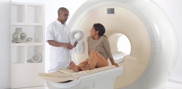
Most ultrasound scans take between 15 minutes and 1 hour. It is usually done in the radiology department of a hospital, and is administered by a radiologist or ultrasonographer. There are 3 main types of ultrasound scanning: external, internal and endoscopic, depending on which part of the body is scanned.
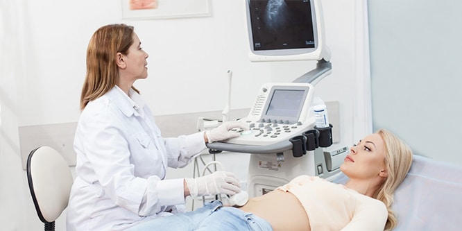
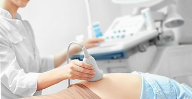
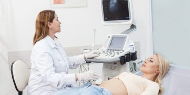
External ultrasound scan
The patient lies on a bed and a water-based gel is applied to the area. The gel helps the probe to come into contact safely with the body, and eliminates any air left between the probe and the skin that may prevent sound waves from entering the body. During the shot, the probe moves over the skin. It is not uncomfortable. After the procedure, the gel is wiped from the skin.
Internal ultrasound scan
Probe is placed inside the body. Internal examination allows a closer examination of organs, such as the prostate gland, ovaries or uterus. During the procedure, it is requested to pull the knees towards the chest, lie on their back or sideways.
A small, sterile ultrasound probe is slowly inserted into the vagina or rectum, and the images are transmitted to the monitor. Internal examinations can be uncomfortable, but usually do not cause any pain, and do not last too long.
Endoscopic ultrasound scan
Probe is attached to a long, thin, flexible tube (an endoscope), and transported further into the body such as the stomach or esophagus. Patient is asked to lie down. Since endoscopic ultrasound scanning can be uncomfortable, patients are usually given sedatives and local anesthetic spray is used to numb the throat. The mouth protector may also be used to protect the patient’s teeth if the mouth is kept open.
If sedatives are given in endoscopic ultrasound, it is recommended to stay in the hospital for a few hours after the procedure. It is necessary to accompany the patient for 24 hours. During this time, patient should not use tools, machinery, or drink alcohol.
Things to do before ultrasound
Although most ultrasound examinations do not require preparation, there are a few exceptions:
- For some scans, such as gallbladder ultrasound, the patient may be asked not to eat or drink anything six hours before.
- A full bladder may be required for examinations such as pelvic ultrasound. The patient may be asked to drink six glasses of water two hours before the screening and not urinate until the screening is finished.
- To improve image clarity, injection of a harmless substance called contrast agent is given before scanning.
- During ultrasound, the patient is asked to take off their accessories. Therefore, it is better to leave valuables at home and wear comfortable clothes.
- The patient may be asked to take off some or all of his/her clothes.
- If necessary, intravenous sedatives may be given.
- Small children may need additional preparations for ultrasound scans.
- In most ultrasound examinations, patients lie on a movable examination table. They may be asked to change positions to improve image quality.
What is a Level 2 ultrasound used for?
It is a kind of ultrasound obtained at the 18th and 23th gestational weeks in which the baby’s organ development is completed and amniotic fluid increases. Level 2 ultrasound performed by perinatologists examines the baby’s development and anatomy in detail.
Detailed ultrasound thoroughly examines the skeletal system of the baby, the structures inside the skull (brain, cerebellum, cerebrospinal fluid channels), facial area, organs and vessels in the rib cage, and organs in the abdomen. It can check whether the placenta is close to the birth canal. The umbilical cord and the volume of amniotic fluid are examined.
Cervical length is measured to predict a possible premature birth risk. It can check whether there is any sign of birth defects or chromosomal disorders. Causes of complications are investigated. Anomalies in uterus can be detected in 70-80% of the cases in a level 2 ultrasound.
What is color Doppler ultrasound used for?
Color Doppler, also known as color ultrasound, uses a computer that converts measurements into a series of colors to show the speed and direction of blood flow through a blood vessel. It checks the flow in the uterine artery that goes to the baby from the right and left side of the uterus. Thus, the likelihood of pregnancy poisoning is assessed. It is usually applied to mothers with developmental problems and some systemic diseases.
It is used to detect any decrease in amniotic fluid or delayed growth. Color ultrasound works well for testicular ultrasound. This high-quality color imaging has made testicular ultrasound a definitive and specific examination.
What is 4D ultrasound, what is it used for?
Two-dimensional (2D) ultrasound image is the most common one. 3D and 4D ultrasounds are possible thanks to improvements in computerized analysis of sound waves recorded from different angles. Three-dimensional images are collected from sound waves that come back from different angles. Images are clearer and more detailed. The difference between 3D and 4D ultrasound is that 4D is similar to a video showing a three-dimensional movement in real time.
Uses and types of ultrasound
Gynecological ultrasound
It monitors lower abdomen, uterus and ovaries, which is done in two ways: abdominal and vaginal. The bladder must be full for it. In vaginal ultrasound, bladder does not need to be full, and it gives a better view when it is empty. Organs are more clearly visualized by a vaginal ultrasound. Egg development capacity, ovary, cysts and tumors can be detected.
Urological ultrasound
It doesn’t require being on an empty stomach or preparing the bowels. The patient may be asked to drink a glass of water an hour before the scan. Ultrasound is performed when lying on the back. The probe is placed between the belly and the pelvic bone. The image is seen on a monitor.
Discharging the bladder is requested in order to check how the bladder is discharged, and the discharged bladder is monitored again.
Prostate (transrectal) ultrasound
Prostate ultrasound which gives a detailed outlook of the prostate gland, is usually performed by placing a special ultrasound probe in the male rectum. The prostate gland and its nearby tissues are examined in the diagnosis of prostate disease. Early detection of prostate cancer requires ultrasound as well as a tissue sample taken from the prostate.
The patient may be asked to wear a gown and to lie down on his/her side by pulling the knees towards the chest. A finger-sized ultrasound probe is inserted into the rectum. If a biopsy is planned, it may be required to stop aspirin and other blood thinners 7 to 10 days before the procedure and and an enema may be needed to cleanse the intestines.
Testicular (Scrotum) ultrasound
Testicular ultrasound helps to evaluate almost all problems in the scrotum, the sac containing the testicles. No preparation is required. The examination is done while lying on the back on the table. The scrotum is put on a towel and warm gel is put on it for transmitting the sound waves and the probe is moved over the area. A well-performed Scrotal Color Doppler Ultrasonography scan takes at least 15 minutes.
Brain (Cranial) ultrasound
Head and Transcranial Doppler are ultrasound examinations in order to evaluate brain tissue and blood flow to the brain, respectively.
Head Doppler
A head ultrasound examination produces images of the cerebrospinal fluid in the brain and the fluid-filled cavities (ventricles) in the deep part of the brain. This examination is usually performed on babies whose skull formation is not completed.
Gaps between the bones of the skull serve as a “window” that allows ultrasound waves to enter and return freely to the brain. Ultrasound probe and some gel are placed in areas where there is no bone. Trans-fontanel ultrasound evaluates the brain development of the baby. This procedure requires no special preparation.
Transcranial Doppler
Transcranial Doppler (TCD) evaluates both direction and velocity of blood flow in the main arteries of the brain. This ultrasound scan is also used during surgical procedures. TCD can be used alone or in other diagnostic examinations such as magnetic resonance imaging (MRI), magnetic resonance angiography (MRA) and computed tomography (CT) scans.
The patient may be asked not to use nicotine-based products that can cause blood vessels to contract before brain ultrasound.
Urinary tract ultrasound
For kidney and urinary ultrasound, the patient lies on his/her back, on his/her tummy and sideways, respectively. The ultrasound probe is brought into contact with the body at different angles. Gel is applied to the skin surface. Bladder must be full during the lower abdomen sonography (pelvic ultrasonography), where the prostate and bladder are evaluated. There is no need to fast when only lower abdominal examination is asked.
Complete abdominal ultrasound
Abdominal USG examines all organs in the abdomen. Abdominal USG monitors the kidneys, liver, gallbladder and pathways, spleen, abdominal aorta, pancreas and other abdominal vessels.
The patient may be asked to eat a lean meal or not to eat 8 to 12 hours before the examination in order to monitor liver, gall bladder, spleen, pancreas and aorta better, and to prevent the accumulation of gas in the intestines.
For ultrasound of the kidneys the bladder has to be full, the patient may be asked to drink 4 to 6 glasses of liquid about one hour before the examination.
Side effects of ultrasound
- Most ultrasound scans are not invasive.
- No radiation is used for ultrasound imaging.
- It is a safe imaging method for the follow-up of babies during pregnancy. Nevertheless, ultrasound is recommended during pregnancy only when medically necessary.
- Those who are allergic to latex should inform the sonographer or physician who performs the scan. In this case, a latex-free probe cover is used.
- Probe may cause discomfort when pressed into the skin or when entering the body.
- Endoscopic ultrasounds can be a bit more uncomfortable, and can cause temporary side effects, such as sore throat or bloating. There is also a risk of internal bleeding in some cases.