ECG, or electrocardiogram, is the process of recording and analyzing electrical activity produced by heart cells that enables the heart to function. It is a simple, short and painless test, and provides important information about the heart’s health. It is a useful examination for the diagnosis of many heart diseases such as heart attack, heart rhythm disorder, and electrolyte disorder. ECG is taken by the electrocardiography device, adhering to the chest front wall, both hands and ankles. Electrocardiographic report is interpreted by a doctor. Electrocardiogram has an important role in the diagnosis of emergency heart diseases, which are manifested by complaints such as chest pain, palpitation and fainting, which are common reasons for emergency services.
Table of Contents
What is electrocardiogram (EKG)?
The heart generates a self-electrical stimulus; this message is transmitted to all heart cells. Heart beats occur in this way. An electrocardiogram is a recording of all the electrical activities produced by the heart cells during the test period.
Why is ECG done?
Electrocardiogram is not a definitive diagnostic method that can be used to diagnose each heart disease. However, it helps to make a diagnosis in line with the complaints of the patient and the symptoms detected after a physical examination as well as the results of laboratory tests.
- To diagnose patients with heart attack symptoms
- To check the functioning of the pacemaker
- For identify the effect of the deficiency and excess of the electrolytes such as potassium and calcium on the heart
- To detect heart rhythm disorders.

Who needs ECG?
- People who has chest pain or shortness of breath
- Patients who experience rapid, irregular beats in the heart
- Those with palpitations
- Patients with abnormal sound heard during examination of the heart with a stethoscope
- People who suffer from fainting and dizziness
- Those who have a pacemaker
What diseases can ECG detect?
- Heart attack: Changes in electrocardiography results occurring due to a heart attack are very important in diagnosis.
- Pulmonary embolism
- Atrial fibrillation
- Heart rhythm disorders
- Hypertrophic cardiomyopathy (a disease characterized by thickening of the heart muscle)
- Potassium, calcium deficiency or excess
- Cardiac arrest
- Stroke: The clot leading to a stroke can be caused by heart rhythm disorder called atrial fibrillation.
ECG types
- Routine ECG: This type of electrocardiography is used routinely. It is widely used in clinics, polyclinics, emergency services.
- Holter ECG (Ambulatory ECG): Routine ECG recording is short. An arrhythmia that exists in other times may not appear during routine ECG. In such cases, heart rhythm is recorded continuously for 24-48 hours if the suspicion of arrhythmia is high. The Holter device is a small device in the size of a mobile phone. The device is used by putting around neck or on the waistband. Recording MRI is not suitable while carrying the device. Devices such as mobile phones and remote controls should not be kept near patient. Obviously, the patient should not take a bath while carrying the device.
- Effort Test (Stress ECG, Stress Test): An electrocardiogram is recorded, if the patient is physically active on the treadmill or the ergo-cycle. In this way, the effects of an effort on the heart can be examined.
Preparations before ECG
Medical history, complaints, symptoms, physical examination and laboratory tests are also examined at the same time as the electrocardiography is planned by the doctor. As a result of all these medical examinations, the cardiologist asks for electrocardiography if it is necessary.
Electrocardiography should be urgently carried out, as it is the case in patients who have symptoms of heart attack. In such cases, there is no preliminary preparation.

How to prepare for an ECG?
- When electrocardiography is performed, conductive electrodes will be attached to the front wall of the chest and to the hands and ankles. Wear comfortable clothes that can be easily taken off.
- Excessive exercise should be avoided before the electrocardiography is carried out. In relation to exercise or excessive physical activity, heart rate is accelerated, resulting in false results.
- Smoking should not be allowed before recording.
- If there is perspiration in places where electrodes are to be attached, they should be dried.
- Sometimes it is difficult for men to attach the electrodes to the chest area due to excessive hair and may cause problems in the electrocardiography. If this is the case, it would be recommended to have a chest shave.
- Medications used before the stress ECG test, and diseases related to heart and other organs should be mentioned to the physician requesting the test.
How is an ECG done?
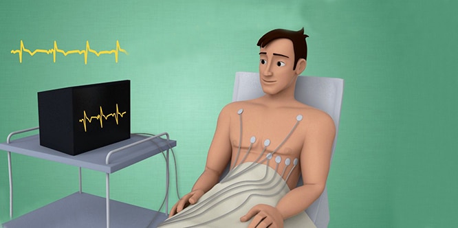
- The person lies on back while the thoracic anterior (front chest) wall with the wrists and ankles remains open. He/she should be comfortable to avoid muscle contraction.
- In order to facilitate the electric current in the skin, a thin layer of gel is applied to the body areas where the electrodes are placed.
- Electrodes are placed on the right and left wrists and ankles. 6 electrodes are placed in certain areas on the thoracic anterior wall.
- Electrocardiography recording is started.
- The patient should not move during electrocardiography and should not talk.
- After recording, the electrodes are removed. The gel is wiped with a towel.
How long does ECG take?
Insertion of electrodes, ECG recording and removal of the electrodes take approximately 10 minutes. This may be longer if the process should be repeated in the case that the patient moves, talks, or has problems with device connections.
ECG results
- The routine ECG test is printed on paper through the ECG device and is then delivered to the doctor. The physician who wants the electrocardiography test can immediately interpret the result.
- The Holter ECG test should be reported by the cardiologist after the end of the test, and this process is longer than the routine ECG.
- After the effort ECG test is completed, the cardiologist will check whether there is an exercise-induced change in electrocardiography results.
- The proper treatment such as drug therapy, angio, and pacemaker replacement is planned as a result of electrocardiography tests.
How to read ECG results?
It is evaluated by the cardiologist after the recording is printed on paper. Routine ECG test is a test which can be evaluated by non-cardiologist doctors when it is taken in the emergencies and the intensive care units.
ECG terms
- Myocardial infarction: Lack of oxygen in the heart tissue of the patient means that he/she has a heart attack. If patients arrive in emergency service with complaints such as chest pain and fainting, electrocardiography is immediately performed. If electrocardiography shows symptoms related to angina or heart attack, urgent intervention is required.
- Heart rhythm disorder: It means that the heart of the patient has extra, irregular beats that should not be there. When severe rhythm disorders are encountered on electrocardiography, immediate intervention is required.
- Heart rate changes (tachycardia, bradycardia): It means that the heart rate is either insufficient or excessive while it should be in the range of 60 to 100 beats per minute.
- Electrolyte imbalance: Elements such as potassium and calcium play an important role in the contraction of the heart muscle and the transmission of the electric current in the heart. Their deficits and redundancies affect the functioning of the heart, and therefore changes occur in the electrocardiogram.
- Side effects of medications: Medications such as beta blockers, calcium channel blockers, digoxin may affect heart rhythm and may cause changes in electrocardiography.
Risks or side effects of ECG
ECG is a simple and painless test. No electrical current passes from the device to the patient. It is safe in this respect. A skin reaction may develop if patient is allergic to gel used during routine ECG, but this is usually a temporary condition. During the stress ECG, the patient may develop complications such as chest pain, shortness of breath, palpitations, dizziness, and fainting.
However, these complaints are not related to the electrocardiography test, but to the fact that the exercise triggers the damage that is already present in the heart.
When can ECG not be done?
There is no obstacle to routine ECG. Stress ECG should not be carried out if the patient has problems such as chest pain, shortness of breath, palpitations immediately before the procedure. In addition, stress ECG should not be done in patients with heart failure, severe aortic valve stenosis, very high blood pressure or severe rhythm disorders.
What are the complaints after ECG?
After the routine ECG or Holter ECG extraction, the patient does not have any complaints. They are painless tests that do not affect anybody’s daily life. Complications such as chest pain, shortness of breath and palpitation may occur in the person after performing a stress ECG test. If these complications start during the test, the tester should be informed immediately. The test will be terminated if necessary.
Suggestions for those who will have ECG
- If you have cardiac problems, and if you have had a previous ECG, take it with you to the cardiologist. It is important to diagnose whether some electrocardiographic changes are already present.
- Always inform your doctor about any other medicines you use and other diseases you have before the stress ECG test.
- Inform your doctor if you have problems such as chest pain, dizziness, shortness of breath during or after an effort ECG test.
- The electrodes used for the Holter ECG device to record can sometimes be dislodged over a 24-hour period, which prevents recording. From time to time you can check if the electrodes are in place and make the necessary intervention.