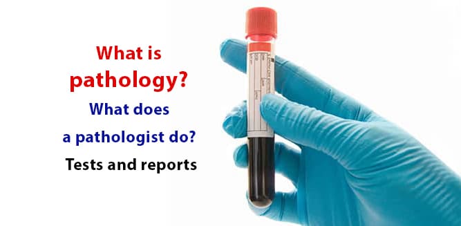
Pathology means the study of disease; it detects changes occurred in tissues and cells by using special methods. A pathologist evaluates the samples taken for diagnosis and writes reports indicating the diagnosis. Pathology tests are often done to diagnose cancer and determine the stage. However, all cells and tissues in the body can be examined by pathology tests, non-cancer diseases can be diagnosed as well. Pathology reports indicates the diagnosis of the pathologist. The stage that the disease has reached is written down. If a diagnosis cannot be identified due to some circumstances, additional examinations or taking new samples may be requested.
Table of Contents
What is pathology?
Pathology consists of the words ‘pathos’ and ‘logos’. ‘Pathos’ in ancient Greek means disease, ‘logos’ knowledge. Pathology, which means ‘the study of disease’, is a specialty in medicine. Organs, tissues and cells in our body have their own unique outlook. Their macroscopic and microscopic images are identified. Many changes occur in the cells, tissues and organs during a disease. Pathology specialists diagnose diseases by examining the changes with specific techniques and tools.
Branches of pathology
- General pathology: It investigates changes in all cells and tissues that occur during the deterioration of the proper functioning of the regular physiological processes. It is concerned with issues such as inflammation, tumor, metastasis.
- Special pathology (system pathology): It examines diseases of the systems and the organs that make up those systems. It can be divided into sub-groups such as lung pathology, neuropathology (examines nerve diseases), gynecopathology (examines cervical and ovarian diseases), dermatopathology (examines skin diseases).
Who is a pathologist?
Following the basic medical education of 6 years, physicians who want to become pathologists pursue a further 4 years of specialization. The pathologist diagnoses diseases by examining samples taken from tissues and organs that are considered to be sick. He/she conducts this examination either with a naked eye or under microscope and uses special techniques when necessary. He/she prepares the pathology report as a result of the investigations.
Areas of study of pathology
The main area of study of the pathology is to undertake the necessary investigations for definitive diagnosis of all kinds of diseases. However, there are also other areas of work:
- Investigation of diseases in animals and testing of treatment methods
- Autopsy examination
- Conducting clinical and pathological investigations with biochemical and microbiological samples taken from the patient
- Genetic examinations

Pathology tests
What is a biopsy?
An extraction of sample cells or tissues for a pathological examination is called a biopsy. A biopsy can be taken from each organ in the body, including the brain. Biopsies can be performed by local anesthesia or during surgery and biopsy samples can be taken on the organs directly through the use of a needle, through endoscopy with the stomach and esophagus, through colonoscopy with the intestines or through bronchoscopy with the lungs. There are 2 types of biopsies:
- Excisional biopsy: the complete removal of the diseased area to be examined.
- Incisional biopsy: the partial removal of the diseased area to be examined.
In some types of cancer, the organ and all the surrounding lymph nodes can be removed and sent to the pathology laboratory.
What is frozen?
In cancer, the organ or surrounding tissues taken during surgery is sent to the pathology laboratory for a rapid examination. Frozen is the preliminary examination done quickly. If a diseased tissue is found after this procedure, surgery is done more extensively. If the examination result is normal, the operation is terminated. Therefore, frozen examination is very important.
What is an autopsy?
In order to determine the actual cause of death, tissues or organs taken from the deceased are sent to the pathology laboratory.
Pathology and cytology
Cytology is made up of the words ‘cyto’ and ‘logos’, which means study of cells. Cytology, due to the fact that disorders at the cellular level are the essential cause of diseases, examines deviations apart from the normal appearance of the cell. Samples are taken from body cavities, secretions and organs, and spread onto a thin glass called lam. Preparations are stained with cytological dyes. Cells are examined under microscope according to their structure, shape and staining properties. Diagnosis is made after evaluating cell conditions that stray from normal appearance and functioning.
Especially in the early diagnosis of cancer, in identifying the response to hormone therapy in some cancers, it serves as an easy and important diagnostical tool. Examined materials are obtained by exfoliative cytology and fine needle aspiration methods.
- Exfoliative cytology: As a result of the renewal of the tissues, the cells fall off from the surface. The method of collecting these fallen-off cells and examining them is called exfoliative cytology. The cells of the female reproductive system such as uterus, cervical organs, the cells of the lower respiratory tract or in the membranes surrounding the organs such as the heart, lung, joint, cerebrospinal fluid are examined by this method.
- Fine needle aspiration cytology: Extraction of fluid through the injection needle after entering into any mass is called aspiration. Cells from samples taken from any mass occurred in external or internal organs can be examined by fine needle aspiration cytology.
What is done in the pathology laboratory?
- The parts taken for pathology examination are sent to the pathology laboratory after being placed in protective fluid to prevent any deterioration.
- Specimens reaching to the laboratory are registered, first their outlooks (macroscopy) are evaluated and the parts considered necessary to be examined by microscope are selected.
- These parts are sampled and undergo processes (tissue tracing) that allow very thin sections to be taken.
- The prepared sections are marked with a special dye and examined in a light microscope.
- If the light microscope does not provide sufficient magnification, the electron microscope can be used.
- Samples taken in the pathology laboratory in order to facilitate the diagnosis can be stained with special dyes and genetic examination can be done.
For which diseases can pathology tests be made?
Pathologic examination is necessary for definitive diagnosis, follow-ups, and treatment responses of many diseases, of which cancer is the prominent beneficiary. Definitive diagnosis in all organ cancers is made by pathology. As with cervix cancer, pathological examination is necessary to diagnose stages which cancer development has reached in order to take the necessary steps.
Pathological examination may also be necessary for the diagnosis of non-cancerous diseases. These include:
- Parenchymal diseases of the lung
- Some intestinal diseases such as ulcerative colitis, Crohn’s disease
- Some skin diseases such as psoriasis
- Thyroid diseases
- Gastric diseases such as gastritis and ulcers
- Liver diseases such as hepatitis
- Infectious diseases caused by virus, bacteria, parasites, fungi
https://www.healthandmedicine.net/what-is-psoriasis-symptoms-causes-and-treatment/
What is a pathology report?
Tissues/organs/cell mass/fluids sent to the laboratory for examination by the pathologist are called specimens. A pathology report is issued to report the results of these samples.
Reading a pathology report
A pathology report should include:
- Name, surname and age of the patient
- Date of the specimen
- Number and date of the report
- Name of the pathologist who writes the report
- Name of the sample examined
- From which part of the body it is taken and by which method
- Dye used when examining the sample
- Which method is applied
- Preliminary diagnosis: It is a report about the suspected disease written by the physician who sends the sample.
Apart from these, a pathology report may include:
- Macroscopic: The visible features of the sample examined, for example, number, color, dimensions of the samples are written in this part of the report.
- Microscopy: Properties of the sample examined under the microscope are described in this part. Information given in this section can only be understood by the physician.
- Diagnosis: Diagnosis of the pathologist. Severity and scale of the findings of the diagnosed disease are indicated. If there are no signs of disease in the examined sample, it may be a “normal finding”. A statement such as “repeating biopsy is recommended” may be included in the report. Diagnosis section of the pathology report gives sufficient information to the doctor who demands the examination in the first place. It becomes clear whether the disease has been found. The treatment method is determined according to the stage of the disease.
Does pathology results give definitive diagnosis?
When all conditions are appropriate, a definitive diagnosis of the disease is possible. These conditions are:
- Sample should be taken from the right place with an appropriate method.
- The material received should be sufficient for examination.
- Material should be examined with a correct method.
- The pathological features of the disease should be specific to that disease.
Sometimes findings of the examination may suggest more than one disease or condition. Or, the material may not provide sufficient information due to the method of extraction of the sample. In such cases, before any definitive diagnosis is made, pathologist may ask to repeat the procedure to obtain additional information or to take a sample with a different method, and after the investigation is completed, a definitive diagnosis is made.
What does “no pathological findings” mean?
In the samples sent to the pathology department, the statement which no pathological finding was found is made in the absence of any diseased tissue or disorder. Unable to observe any pathological finding does not always indicate that the person is healthy. This expression is used for the sample taken. Specimens may be taken from intact tissue and cells. In such cases, sampling is repeated if the presence of the disease is still suspected.
What does the delay of pathology result mean?
After the material to be examined reaches the pathology laboratory, the time it will take to write the report is not fixed. This period may be prolonged if the material is taken from a hard-to-examine tissue (such as a bone), if there is a need for a special procedure for the diagnosis of the material being investigated, or there is inadequacy of staff in the laboratory due to the intensity of the workload.
Therefore, if there is a delay in publishing of the report, it should not be considered a result of negligence. Cytological examination reports are prepared in a shorter time than other examinations. In some cases, pathology results may take a full month to be reached.
Can samples be meddled in a pathology laboratory?
Samples are sent to the laboratory with a uniquely barcoded form, including information about the patient and the sample taken. Once the sample is delivered to the laboratory, the number is given, registered and labeled. Names on the container and in the form are verified by the pathologist before examination. Precautions are taken to avoid mistakes such as tissue, preparation, and/or report meddling. If you think you are in such a case, you should talk to your doctor. The doctor may contact the laboratory if necessary. For more>>> Laboratory Tests


