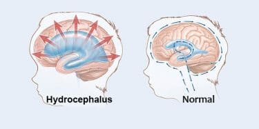
Hydrocephalus is a chronic and neurological disease caused by the over-accumulation of cerebrospinal fluid. It can be seen at any age, including newborns. Pressure inside the skull causes many problems. Potential brain damage occurring as a result of fluid accumulation may cause developmental, physical and mental deterioration. In order to avoid serious complications, treatment is required. Since hydrocephalus does not recover spontaneously and cannot be treated with medication, the only solution is surgery. Shunt-mounted surgery should be performed as soon as possible after the disease is diagnosed. Most treated patients can resume a normal life. Without treatment, death may occur.
Table of Contents
What is hydrocephalus?
In hydrocephalus, which means “water filled head” in Greek, the cerebrospinal fluid accumulates excessively inside the skull and this causes pressure on the brain. This disease, which can be seen from the mother’s womb until later ages with the help of radiological techniques, manifests itself with different signs and symptoms in each age range.
Early diagnosis and treatment is very important. When treatment is delayed, irreversible brain tissue damage and permanent neurological disorders can occur.
Causes of hydrocephalus
The fluid, which circulates between spaces in the brain called the ventricle and surrounds the brain and spinal cord, is called “cerebrospinal fluid”. This fluid acts as a buffer against the transmission of nutrients, disposal of wastes and impacts from the outside world. This is an important element for balancing pressure changes in the brain and spinal cord.
This fluid is continuously in the state of production, circulation and absorption. The increase in the production of this fluid, the disorder in the circulation (stenosis or obstruction in the ducts) and the disturbances in the absorption can cause the increase in the amount and pressure of the fluid, enlargement of the brain spaces in which it is contained, damage to the brain tissue cells, and this condition is called hydrocephalus.
Causes of congenital hydrocephalus
The cause of some congenital hydrocephalus cases is not known exactly. However, if a baby is born with hydrocephalus, it may be a brain problem limiting the flow of cerebrospinal fluid (CSF). Congenital hydrocephalus can also be seen in premature infants. Some premature babies suffer from brain hemorrhage. As a result, CSF flow is prevented and hydrocephalus occurs. Other possible causes of congenital hydrocephalus:
- Some conditions such as spina bifida
- Mutation of X chromosome
- Rare genetic disorders such as Dandy Walker malformation
- Fluid-filled sacs between the brain or spinal cord and the arachnoid membrane

Causes of acquired hydrocephalus (in children and adults)
Hydrocephalus that develops in adults and children is usually caused by a disease or injury that affects the brain. The possible causes of this condition called acquired hydrocephalus include:
- Bleeding in the brain (eg. blood leakage from the surface of the brain as in subarachnoid hemorrhage)
- Blood clots in the brain (venous thrombosis)
- Meningitis (infection of membranes surrounding the brain and spinal cord)
- Brain tumors
- Head injury
- Stroke
In some people, there may be congenital stenosis in the passages through which the cerebrospinal fluid flows, but symptoms may not appear for years.
Causes of normal pressure hydrocephalus
Hydrocephalus that develops in the elderly may occur after brain damage, bleeding in the brain or after infection. Or it may be linked to other health conditions that affect normal blood flow, such as diabetes, heart disease or high cholesterol. However, in most cases, there is no definite cause.
Symptoms of hydrocephalus
Hydrocephalus or fluid in the brain causes different symptoms depending on the type of hydrocephalus and the age of the affected person.
Symptoms of congenital hydrocephalus
- The head is larger than normal,
- Fine and shiny scalp, where veins are easily visible
- Slippage of the eyes or the tightness of the fontanel (soft area above the baby’s head)
- Nutritional disorder
- Irritability
- Vomiting
- Sleepiness
- Muscle stiffness and spasms in the lower extremities
Congenital hydrocephalus can sometimes be detected before the baby is born during ultrasound screening. However, it is usually diagnosed immediately after birth during newborn physical examination. The most important symptom is that the baby’s head is larger than normal.
Symptoms of acquired hydrocephalus
- Headache
- Neck pain
- Feeling sick
- Sleepiness (possibility of coma)
- Mental turbidity and confusion
- Blurred or double vision
- Difficulty in walking
- Urinary incontinence and in some cases bowel incontinence
Headaches may get worse in the morning because the brain fluid can accumulate during the night as it does not discharge well when it is in the extended position. Also, sitting for a while can also increase headache. However, as the condition progresses, headaches may become permanent.
Symptoms of normal pressure hydrocephalus
One of the first signs of normal pressure hydrocephalus, which is more common in adults over 60 years of age, is to fall suddenly without losing consciousness. Other symptoms include:
- Gait changes and gait disturbance
- Mental function deterioration, such as memory problems
- Urinary and bowel incontinence
- Headache
Diagnosis of hydrocephalus
With the advancement of neuroradiological techniques, magnetic resonance imaging (MRI) and computed tomography (CT) can be used to detect changes in brain cavities and changes in brain tissue. MRI and ultrasonography performed in the womb in prenatal period are used to detect changes in the brain spaces and follow the brain development of the baby. With high-resolution MRI after birth, more information about the cause of hydrocephalus can be obtained.
Treatment of hydrocephalus
Is there any drug treatment for hydrocephalus?
Today, the drug treatment of hydrocephalus is not possible. Surgical intervention is the only treatment method.
Hydrocephalus shunt surgery
The most common method in the treatment of hydrocephalus is shunt surgery. In this operation, a drainage system called shunt is placed in the brain. Shunt is a long flexible tube with a valve that allows the fluid in the brain to flow in the right direction and at the appropriate rate.
One end of the tube is usually inserted into one of the ventricles of the brain and the other in a space of the abdomen or heart where the excess cerebrospinal fluid is more easily absorbed. Surgery is performed under general anesthesia. Patients with hydrocephalus often need this shunt system for their survival. They can continue their lives without any problem with the help of regular monitoring.
Endoscopic third ventriculostomy
Endoscopic third ventriculostomy is a surgical procedure that can be performed for certain people. In the procedure, the surgeon uses a small video camera (neuroendoscope) that uses fiber optic technology to look directly into the brain. It drills a small hole at the bottom of the third ventricle with a small instrument. Thus, the cerebrospinal fluid can overcome the obstruction and flow towards the absorption site around the surface of the brain.
Hydrocephalus surgery in infants
The first step is to ensure that the baby is born as early as possible when the diagnosis is made in the womb. The surgery is performed immediately. Shunt surgery and endoscopic third ventriculostomy are also the methods that can be applied to infants.
Outcomes after shunt surgery and risks
Shunt tools are not perfect. Regular follow-up of the patient is very important after shunt surgery. Some complications may develop after surgery. In case of any complication, re-operation may be necessary to replace the defective part or the entire shunt system. For example, 50% of shunt surgeries in children should be repeated in two years.
The most common shunt complications are shunt failure and infection:
- Shunt failure: Shunt failure is usually caused by clogging. When occlusion occurs, the cerebrospinal fluid that cannot be discharged accumulates and the symptoms of hydrocephalus reappear. Clogging is often caused by blood cells, tissues or bacteria. Apart from this, although the shunts are very durable, sometimes some components may crack or break as the child grows. Occasionally, they may come out of place or, in rare cases, the valve may malfunction.
- Shunt infection: Shunt infection is often caused by the patient’s own bacterial organisms. The most common infection is Staphylococcus Epidermidis, which is normally found on the skin surface and in sweat glands and hair follicles. This type of infection can usually be seen one to three months after surgery. A shunt-induced infection may cause symptoms such as mild fever, pain in the neck or shoulder muscles, and redness or tenderness along the shunt path in addition to common symptoms of hydrocephalus.
If you suspect infection, it is very important that you tell your neurosurgeon immediately or go to the emergency room. Shunt infections are serious and require immediate medical attention to avoid life-threatening diseases or possible brain damage.
Other shunt complications
- Excessive drainage: Sometimes the shunt can drain the cerebrospinal fluid faster than it is produced. This is a serious problem. This problem may cause collapse of the ventricles, rupture of blood vessels and headache, bleeding or cleft ventricular syndrome.
- Low drainage: When the cerebrospinal fluid is not evacuated quickly enough, the symptoms of hydrocephalus begin to reappear. If the shunt has a regulated pressure valve, this discharging problem can be solved by means of a special magnet placed on the top of the scalp, facing the top of the valve.
- Subdural hematoma: It is a condition where blood is trapped between the brain and the skull as a result of a rupture of blood vessels. It is seen in the elderly and can be treated with surgery.
If you suspect that the shunt system is not working properly, for example if the signs of hydrocephalus have restarted, you should seek immediate medical attention.
How long do hydrocephalus patients live?
Most treated hydrocephalus patients live a normal life. However, when untreated, progressive hydrocephalus is usually fatal. In untreated hydrocephalus, death occurs as a result of tonsils herniation due to increased intracranial pressure following compression of the brain stem and respiratory arrest.
Does hydrocephalus go by itself?
Hydrocephalus is not a self-healing disease. When the diagnosis is made, treatment should be initiated as early as possible so that the neurological losses do not become irreversible.
Suggestions for hydrocephalus patients
- Get information about hydrocephalus and its possible complications in order to manage your disease well and notice when there is a problem. In a complex disease such as hydrocephalus, it may sometimes be difficult to distinguish between a routine medical problem and a life-threatening symptom.
- If you are using aspirin and similar blood-thinning drugs, you should stop taking these medications a while before the surgery. Instead, you can request from your doctoranti-coagulant drugs that do not prolong the bleeding time.
- Short-term antibiotics can be used to prevent postoperative infection
- Do not try to operate the shunt by pressing it from the outside by hand, shunt examination should always be done by your doctor.
- Especially, do not lay the baby on the side of the shunt.
- Shunt can be used for many years, if there is no problem, shunt need not be removed.
- Regular and frequent examination is advised to prevent shunt complications.
- MRI and brain tomography can be performed after shunt surgery. If the shunt valve pressure adjustment is external, it is recommended to consult your doctor about whether it is suitable for MRI. For more:>>> Hydrocephalus


